
0%

Fiber optic laryngoscope blades are specialized airway tools designed to improve visibility during intubation. They use integrated optical fibers to transmit light from the handle toward the blade tip. This illumination helps clinicians identify the tongue, epiglottis, and vocal cords in a confined oral space. The design appears simple. Its clinical role is not.

In practical airway management, blade shape, size, curvature, and optical performance can affect handling. Macintosh-style blades usually lift the epiglottis indirectly, while Miller-style blades support more direct elevation. Fiber optic laryngoscope blades may provide brighter, more focused illumination than conventional blades. This can be valuable in operating rooms, emergency departments, ambulances, and teaching environments. However, better lighting cannot replace positioning, suction, patient assessment, or practiced technique.

Material quality also matters. Stainless steel blades offer durability and repeated sterilization potential. Disposable versions can reduce cross-contamination concerns when used according to institutional policies. Clinicians should check the light pathway, connector, locking mechanism, and blade surface before use. A small obstruction may reduce visibility at a critical moment. That detail is easy to overlook.

This article examines how these blades work, where they fit in modern laryngoscopy, and what features deserve careful evaluation. It also considers limitations, including fogging, maintenance demands, battery dependence, and operator learning curves. No device guarantees a successful airway. That remains an important correction to overly confident product claims. Reliable selection depends on clinical experience, manufacturer instructions, verified performance data, and local infection-control requirements.
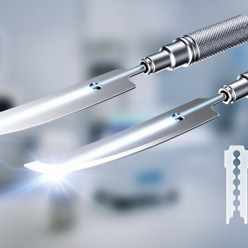
What Are Fiber Optic Laryngoscope Blades?

Definition and Basic Structure of Fiber Optic Laryngoscope Blades

A fiber optic laryngoscope blade is an airway instrument used to expose the larynx during intubation. Unlike a basic illuminated blade, it carries light through a bundle of fine optical fibers. The light travels from the handle connection to the blade tip. This design helps illuminate the mouth, tongue, and epiglottic area in a narrow airway.

The basic structure includes a shaped metal or polymer blade, a proximal connector, an internal fiber bundle, and a distal light window. The blade may have a straight or curved profile. Its flange guides the tongue aside, while the tip supports controlled lifting near the epiglottis. Some models include a smooth channel for guiding the endotracheal tube. Small differences in curvature can affect visibility and handling.

In clinical training, careful inspection matters. The fiber bundle should transmit an even, bright light without dark spots. The tip must remain smooth and securely attached. Any loose joint, cracked surface, or weak illumination can reduce control. Cleaning procedures also require attention, because hidden moisture may damage internal components. The structure seems simple, but it is not completely foolproof. A well-made blade cannot replace positioning, anatomical knowledge, or proper airway assessment. Various patient anatomies may still demand different blade shapes and techniques.

How Fiber Optic Laryngoscope Blades Work

What Are Fiber Optic Laryngoscope Blades?

Fiber optic laryngoscope blades use thin optical fibers to carry light from the handle to the blade tip. This creates focused illumination inside the mouth and throat. The blade itself does not usually transmit the whole image. Instead, the operator views the airway directly along the blade’s path. A bright distal light can reveal the tongue, epiglottis, and vocal cords more clearly than a weak conventional bulb.

How Fiber Optic Laryngoscope Blades Work

During use, a trained clinician guides the blade carefully over the tongue. The curved surface helps move soft tissue without pressing directly on the teeth. As the blade reaches the vallecula or lifts the epiglottis, the light exposes the glottic opening. Small changes in angle often improve the view. Position matters greatly.

A clean optical pathway is essential. Dried secretions, fogging, damaged fibers, or a low battery can reduce brightness. The fibers may also develop dark spots when individual strands fail. That problem is easy to overlook. Blade design, patient anatomy, and operator technique all affect visibility. In practice, fiber optic illumination improves access, but it cannot replace airway assessment or careful handling. I have found that even a well-lit view may remain incomplete when the tongue is large or the airway is unexpectedly angled. Reprocessing instructions, routine inspection, and compatible light sources help maintain reliable performance.

Main Types and Design Features

Fiber optic laryngoscope blades are airway instruments that use an internal light bundle to illuminate the larynx. Unlike older external-light designs, they send light toward the blade tip. This can improve visibility during direct laryngoscopy, especially in dim clinical settings. However, brightness alone does not guarantee a clear view.

The main types are curved Macintosh-style blades and straight Miller-style blades. Curved blades usually sit in the vallecula and lift the epiglottis indirectly. Straight blades pass closer to the epiglottis and may help with small or anatomically different airways. Pediatric sizes are shorter and narrower, with tips designed for delicate oral structures. Fit matters.

Design features influence both control and safety. A smooth, rounded flange can reduce tissue pressure during insertion. A reinforced fiber bundle supports consistent illumination, but damaged fibers may create dark spots. The distal tip should remain visible without blocking the operator’s line of sight. Some blades include anti-fog surfaces or channels for suction and oxygen delivery, depending on the model.

Reusable blades require careful cleaning and inspection between procedures. Single-use blades reduce reprocessing concerns but create more waste. In practice, handle compatibility, blade geometry, and patient anatomy must be assessed together. No blade suits every airway. I have found that a familiar blade can feel safer than a technically advanced one, although personal comfort should never replace clinical training or equipment checks.

Clinical Uses and Compatibility

What Are Fiber Optic Laryngoscope Blades?

Clinical Uses and Compatibility

Fiber optic laryngoscope blades use a light-transmitting fiber bundle to illuminate and view the airway. Unlike standard direct blades, they can improve visualization when the tongue, teeth, or neck position limits the line of sight. Clinicians may use them during routine intubation, anticipated difficult airways, emergency procedures, and some pediatric cases. Small blades matter. A narrow mouth opening may require a slimmer profile and gentler insertion.

Compatibility requires more than a matching handle. The blade must fit the handle’s locking mechanism, light interface, and intended power source. Some systems require specific connectors or adapters. Reusable blades also need validated cleaning and sterilization cycles. A blade that connects successfully may still produce weak illumination or an unstable image. That mistake is easy to miss during a busy airway procedure.

The Fourth National Audit Project reported 133 major airway complications in the United Kingdom, highlighting the value of planning, equipment checks, and backup techniques. Clinical teams should confirm blade size, illumination, image quality, and reprocessing status before use. They should also inspect fibers for damage, clouding, or broken tips. A compatibility chart helps, but it is not a substitute for hands-on testing. In practice, staff may discover that “universal” equipment is not truly universal. Manufacturer instructions and facility protocols should guide final selection.

What Are Fiber Optic Laryngoscope Blades? - Clinical Uses and Compatibility

Blade Type or Feature Typical Sizes Primary Clinical Uses Key Characteristics Compatibility Considerations Important Limitations
Fiber optic Macintosh blade Commonly numbered 2, 3, and 4; pediatric sizes may also be available Routine adult and pediatric orotracheal intubation; emergency airway management; operating-room use Curved blade; tip is placed in the vallecula to indirectly lift the epiglottis; integrated fiber-optic light transmission improves illumination Must match the handle’s blade attachment mechanism, dimensions, and light-source interface; standard-compatible systems may follow ISO 7376, but verification is required May provide less direct control of a floppy epiglottis than a straight blade; difficult airways may require an alternative device
Fiber optic Miller blade Commonly numbered 0, 1, 2, 3, and 4 Neonatal and pediatric intubation; patients with a relatively anterior larynx; situations requiring direct epiglottic control Straight blade; tip is advanced beneath or onto the epiglottis for direct elevation; fiber-optic bundle carries light toward the distal tip Size selection must account for patient age, oral anatomy, and handle compatibility; attachment and illumination standards should be checked before use Requires careful technique to reduce contact with the epiglottis and surrounding tissues; an improperly sized blade can obstruct the view or cause trauma
Standard fiber optic blade Usually available in adult, pediatric, and neonatal ranges General direct laryngoscopy when a reusable blade and compatible handle are available Contains a fiber-optic light bundle rather than a conventional bulb mounted at the distal tip; commonly offers a clear working area around the line of sight Confirm hinge geometry, locking mechanism, handle fit, lamp or LED coupling, and electrical or optical specifications where applicable Light transmission can decline if fibers are damaged, contaminated, or poorly maintained
Reusable fiber optic blade Available across most adult, pediatric, and neonatal blade sizes High-volume clinical settings with validated cleaning, disinfection, or sterilization processes Designed for repeated use; may reduce waste per procedure when correctly reprocessed and maintained The blade must be compatible with the intended handle and with the healthcare facility’s validated reprocessing method; follow the manufacturer’s instructions for use Inadequate cleaning or sterilization may create infection risks; repeated processing can eventually affect joints, finishes, seals, or fiber bundles
Single-use fiber optic blade Commonly offered in neonatal, pediatric, and adult sizes Emergency departments, ambulances, operating rooms, and settings where rapid availability and infection-control convenience are priorities Supplied for one patient or one procedure according to the product labeling; avoids routine reprocessing between patients A disposable blade is not automatically compatible with every reusable handle; confirm the stated handle platform, connector design, and light-source requirements Higher per-procedure material cost and increased clinical waste; do not reuse unless specifically authorized and validated
Fiber-optic illumination system Not size-specific; depends on the blade and handle design Improved illumination during direct laryngoscopy, particularly in reduced ambient light or challenging clinical conditions Light is generated in or near the handle and transmitted through optical fibers to the blade tip, keeping the distal profile relatively unobstructed Compatibility depends on mechanical attachment, optical alignment, light intensity, and the applicable handle-blade standard; ISO 7376 compatibility should not be assumed without confirmation Illumination does not replace correct patient positioning, suction, preoxygenation, or an appropriate difficult-airway plan
Compatibility and selection checklist Select according to patient anatomy and intended procedure Applicable to all direct-laryngoscopy procedures using a fiber optic blade Evaluate blade shape, size, visibility, structural condition, illumination, and ease of insertion before clinical use Check handle type, hinge or locking design, light-source interface, standard compliance, intended reprocessing method, and instructions for use A blade that physically attaches may still provide inadequate illumination or fail infection-control requirements; compatibility should be clinically and technically verified

Note: Fiber optic laryngoscope blades are used with a compatible laryngoscope handle to illuminate and expose the laryngeal inlet during direct laryngoscopy. Size selection and clinical use should be determined by trained healthcare professionals, patient anatomy, airway assessment, and applicable institutional procedures.

Cleaning, Maintenance, and Safety Considerations

What Are Fiber Optic Laryngoscope Blades?

Fiber optic laryngoscope blades use embedded light fibers to illuminate the airway during intubation. Their narrow joints and optical channels can retain blood, mucus, or moisture. The CDC classifies blades contacting mucous membranes as semicritical devices. Therefore, they require sterilization or, at minimum, high-level disinfection.

Cleaning should begin immediately after use. Wear gloves, eye protection, and suitable protective clothing. Remove visible soil with compatible detergent and a soft brush. Do not force brushes through delicate optical channels. The FDA’s 2015 reprocessing report stresses that reusable devices need validated, device-specific instructions. Not every blade tolerates immersion. That detail is often missed.

Follow the manufacturer’s instructions for disassembly, chemical concentration, temperature, and exposure time. AAMI ST79 emphasizes cleaning before disinfection or sterilization, with documented processing records. Where required, perform a leak test before immersion. Dry every surface completely. Trapped droplets can support corrosion and microbial survival. Inspect the tip, hinge, locking mechanism, and fiber bundle under bright light. A cloudy lens or loose joint deserves removal from service. Test illumination and blade movement before patient contact. Test it twice. In practice, a visually clean blade may still hide residue inside a hinge. Staff should review failed cleaning cycles, because routine habits can become unsafe shortcuts. Store processed blades in clean, protected areas, separated from contaminated equipment.

What Are Fiber Optic Laryngoscope Blades?

Cleaning, maintenance, and safety should follow the blade manufacturer’s instructions for use. The chart below presents a typical reusable-blade processing sequence; the sequence position is not a duration or clinical rating.

Safety considerations: Inspect the blade, light bundle, distal tip, and locking mechanism before use. Begin point-of-use treatment promptly after use, clean before disinfection or sterilization, and follow the validated processing method specified for the device. Dry the blade completely before storage, protect the optical surfaces from scratches, and remove any blade with visible damage, impaired illumination, corrosion, or compromised mechanical function.

FAQS

What is a fiber optic laryngoscope blade?

It is an airway instrument used by trained clinicians during intubation. Thin optical fibers carry light from the handle to the blade tip. The light reveals the tongue, epiglottis, and vocal cord area. It does not usually transmit the airway image.

What parts make up the blade?

Common parts include a shaped blade, proximal connector, fiber bundle, and distal light window. The blade may be straight or curved. A flange guides the tongue aside. Some designs include a tube-guiding channel.

How does the fiber optic system improve visibility?

The internal fibers send focused light toward the mouth and throat. A bright tip can clarify tissues inside a narrow airway. Position still matters greatly. More light does not guarantee a complete view.

What are the main blade shapes?

Curved blades usually rest near the vallecula and lift the epiglottis indirectly. Straight blades approach the epiglottis more directly. Shorter, narrower sizes may suit smaller patients. Fit matters.

What should be checked before use?

Inspect the tip, connector, surface, and light output. The light should appear even and bright. Dark spots may indicate damaged optical fibers. Check for cracks or loose joints. Small defects can matter.

Why might the airway view remain poor?

Fogging, secretions, weak batteries, and damaged fibers can reduce illumination. Tongue size and airway angle also affect visibility. A bright view may still be incomplete. Technique and positioning remain important.

How should reusable blades be maintained?

Follow the approved cleaning and reprocessing instructions. Remove moisture from hidden areas. Inspect the blade after cleaning and before storage. Moisture can damage internal components. Cleaning is not foolproof.

How should clinicians choose between blade types?

Selection depends on anatomy, patient size, blade geometry, and clinician experience. Handle compatibility must also be confirmed. A familiar blade may feel easier to control. That feeling should not replace training or equipment checks.

Conclusion

Fiber optic laryngoscope blades are medical instruments designed to help clinicians visualize the larynx during airway management and intubation. They typically consist of a curved or straight blade, an integrated fiber optic light bundle, and a connector that attaches to a compatible handle. The fiber optic system transmits bright illumination to the blade tip, allowing the user to see anatomical structures more clearly, even when direct lighting is limited.

Common designs include curved and straight blades, with variations in size, profile, and tip shape to suit different patients and clinical situations. Fiber optic laryngoscope blades may be used in emergency care, anesthesia, surgery, and other airway procedures, provided they match the appropriate handle and equipment system. Proper cleaning, inspection, sterilization, and routine maintenance are essential for safe performance. Users should check the blade for damage, confirm adequate illumination, and follow approved handling procedures to reduce the risk of cross-contamination and equipment failure.

Evelyn

Evelyn

Evelyn is a highly experienced marketing professional dedicated to helping businesses understand products, industry trends, and practical solutions that support long-term growth. With a strong understanding of the company’s product portfolio, customer needs, and evolving market conditions, she......